Top Tips for Retinal Surgery Recovery
On average there are 28,000 cases of retinal detachment or tears in the USA per year, most of which required surgery for full recovery. Whereas this is not initially a painful condition, it is nonetheless a medical emergency. If left untreated then the end result would be impaired vision, but if repaired, and the patient acts upon the recovery precautions, then a full recovery is expected.
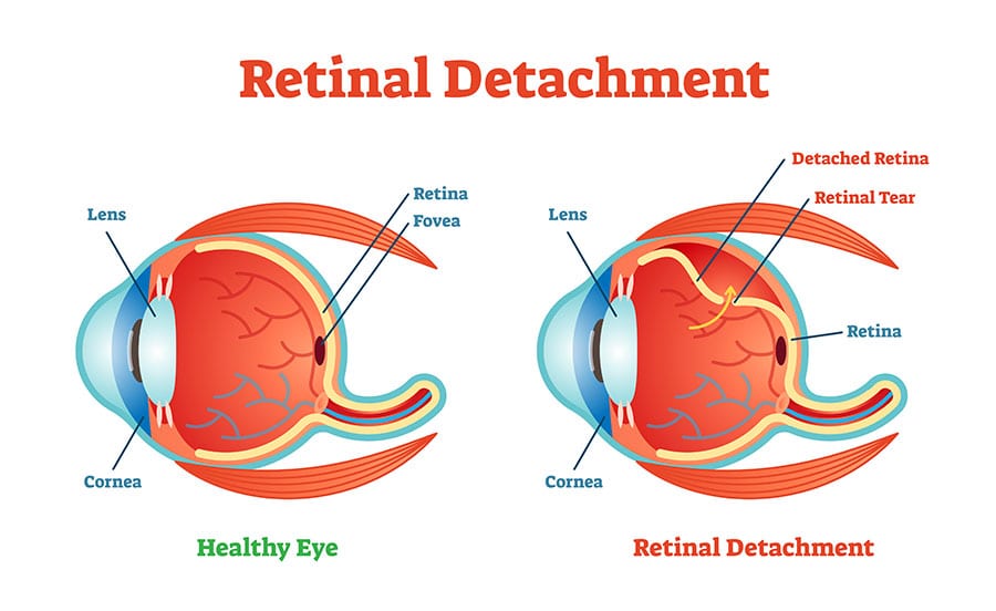
What is the Retina and What is its Function?
The retina is a thin layer of tissue which is located at the back of the eye near the optic nerve. It is light sensitive and receives light from objects we see and converts them into neural signals to send to the brain for interpretation and recognition. These light sensitive cells detect intensity and color which provides our brains with more information so that it can determine what we are looking at. If there is damage to the retina then this will temporarily or permanently impair vision.
There are 3 types of detached retina:
- Rhegmatogenous retinal detachment – this is where a hole, break or tear in the retina allows vitreous fluid into the subretinal space.
- Secondary retinal detachment – this occurs due to an inflammation or vascular abnormality in the eye which causes fluid to collect under the retina.
- Tractional retinal detachment – is when inflammation or injury causes the sensory retina to be pulled away from the retinal pigment epithelium.
These conditions are not only caused by injury to the eye but also occur regularly in patients with diabetes, myopia, severe near-sightedness or who have had cataract surgery. Other eye diseases may also lead to a high incidence of retina detachment which include retinoschisis and lattice degeneration.

How do you Know if Your Retina has Detached?
As leaving damaged retinas untreated can cause long term vision issues, it is important to recognize and act upon symptoms of a detached retina as soon as early signs occur.
These may include:
- An increase in the size and number of floaters.
- Flashes of light (Photopsia) in one or both eyes – these are more likely to occur when the eye is moving.
- A noticeable shadowing in your peripheral vision.
- What looks like a ‘grey curtain’ covering part of your usual vision.
- Blurred vision.
- A sudden decrease in vision.
- Straight lines or objects begin to appear curved.
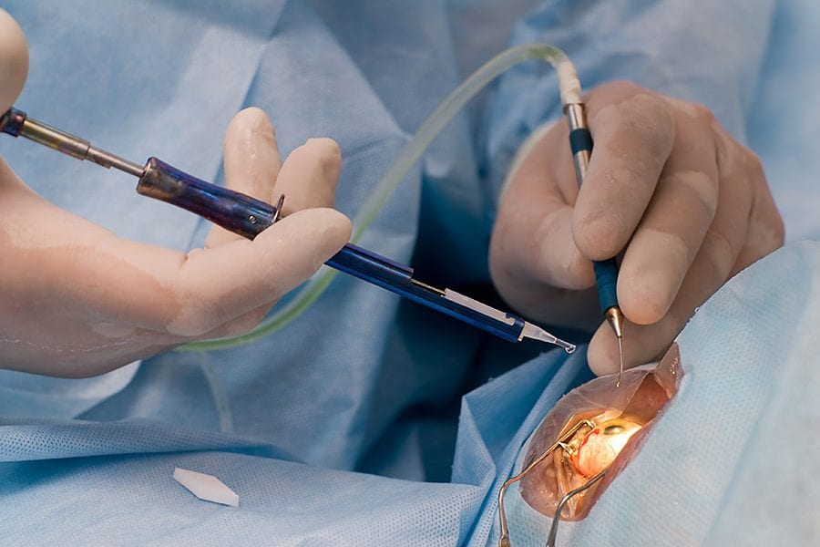
Types of Retinal Surgery
To prevent total sight loss, surgery is usually necessary to repair any tears or leaks. There are several different types of surgery that could be performed:
- Vitrectomy - In this procedure the surgeon extracts the vitreous along with any tissue that is pulling on the retina. A gas or silicone oil bubble is then injected into this area which flattens the retina and holds it in place. If silicone oil is used, then this is usually removed between 2 and 8 months later. If a gas or air bubble is used, this will eventually be absorbed, and no further intervention is necessary.
- Pneumatic retinopexy – this surgery is often used for less complicated detachments. The area where the tear has occurred is frozen (by cryopexy) and a bubble of air or gas is injected into the vitreous cavity. This pushes the retina back against the wall of the eye which stops the flow of fluid into the space behind the retina.
- Scleral Buckle – this procedure involves attaching thin bands of silicone to the sclera area (outside white of the eye). This keeps the retina in place to allow it to reattach.
- Laser Eye Surgery – a laser is used around the tear which creates scar tissue that fuses the retina back in place.

Recovery from Retinal Detachment Surgery
Following retinal surgery your eye will most likely be painful, scratchy or itchy which may last a couple of days. Your surgeon will probably have used some antibiotic ointment or drops in your eye shortly after surgery, to prevent any infections being caused by the operative incisions. You will need to keep using this for a period of time post op. An eye patch or shield is also encouraged to prevent any contaminates from entering the eye and possibly causing new infections.
In addition to antibiotics, you may also be prescribed pain medication to minimize the impact of pain on your rest and recovery period. It is advisable not to lift any heavy objects or move your eyes in a rapid motion as this can also increase the pressure in your eye which could impede recovery. If your eye is swollen then apply an ice pack regularly for short periods of time for the first couple of days (never apply ice directly to the skin, always ensure there is a cloth barrier).
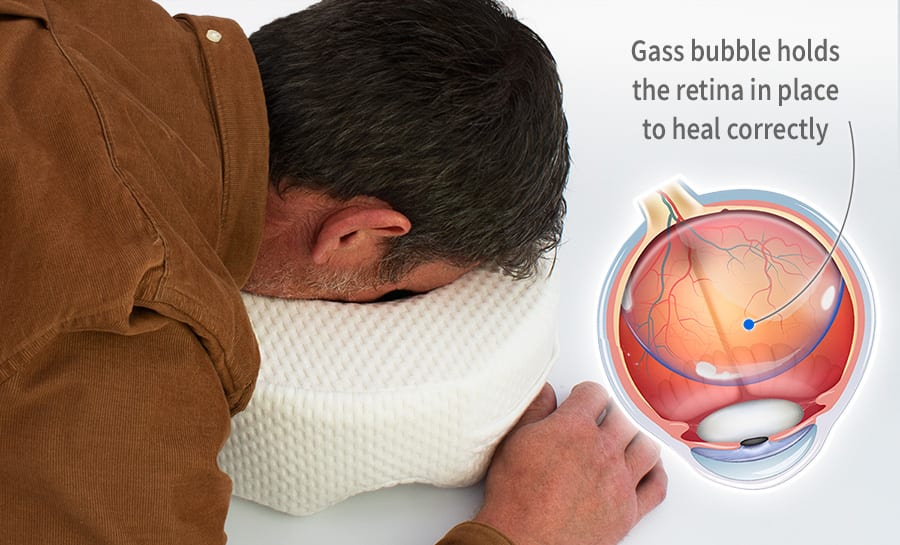
The above advice is standard for most eye procedures, however there are extra precautions that are necessary following retinal detachment surgery due for the need to keep the air/silicone bubble in the correct place. The most important of which is to keep your head in a face down position (parallel to the ground). The AAO (American Academy of Ophthalmology) recommends patients keep their head in this position for 16 hours a day (taking breaks every 15 minutes) for up to 8 days. However, there are a number of practical implications that patients are advised to think about and prepare for before having the surgery:
- Plan to have a family member or friend to support you for the period of time that you need to remain face down. Walking around in this position can be hazardous and recovery does not want to be compromised by a further injury.
- Purchase a Face Down Pillow to make sitting and resting more comfortable. For most people resting face down is an usual position and sitting in this position is certainly unusual. As such there is often new stresses and strains put on the neck, shoulders and back when in this position for long periods of time. Using a good quality face down pillow when sitting or resting can alleviate these and ensure you get the rest you deserve to allow your body to heal. Suitable pillows should be specifically for resting face down, have an extra wide eye socket area to ensure there is no pressure put on the eyes. Large ventilation arches are also recommended to ensure a good flow of air and prevent heat build-up.
- Use mirrors so that you can see objects in front of you. These can be angled so that watching the television or talking to visitors is made easier.
- Avoid flying or scuba diving until the bubble has been absorbed or removed. Changes in altitude cause pressure in the eye to change which can lead to the retina not attaching correctly.
Recovery from retinal surgery can take up to three weeks and it is important to allow yourself the time to recover. Trying to rush the process can lead to further complications so book time off work, relax and give yourself the time to heal. Always seek medical advice if you are concerned or experience unexpected side effects.

Recovering from retinal detachment surgery using a face down pillow to keep the air/silicone bubble in place.
Recovery from retinal surgery can take up to three weeks and it is important to allow yourself the time to recover. Trying to rush the process can lead to further complications so book time off work, relax and give yourself the time to heal. Always seek medical advice if you are concerned or experience unexpected side effects.


Please note: all information, content, and material contained in this article is curated from the sources below and provided for informational purposes only. It is not intended as advice, or as a substitute for the medical advice of a physician. Always seek the guidance of your doctor or other qualified health professional regarding your health or medical condition.